What Is Calcaneal Apophysitis?
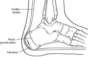
Calcaneal apophysitis is a painful inflammation of the heel’s growth plate. It typically affects children between the ages of 8 and 14 years old, because the heel bone (calcaneus) is not fully developed until at least age 14. Until then, new bone is forming at the growth plate (physis), a weak area located at the back of the heel. When there is too much repetitive stress on the growth plate, inflammation can develop.
Calcaneal apophysitis is also called Sever’s disease, although it is not a true “disease.” It is the most common cause of heel pain in children, and can occur in one or both feet.
Heel pain in children differs from the most common type of heel pain experienced by adults. While heel pain in adults usually subsides after a period of walking, pediatric heel pain generally doesn’t improve in this manner. In fact, walking typically makes the pain worse.
Causes
Overuse and stress on the heel bone through participation in sports is a major cause of calcaneal apophysitis. The heel’s growth plate is sensitive to repeated running and pounding on hard surfaces, resulting in muscle strain and inflamed tissue. For this reason, children and adolescents involved in soccer, track, or basketball are especially vulnerable.
Other potential causes of calcaneal apophysitis include obesity, a tight Achilles tendon, and biomechanical problems such as flatfoot or a high-arched foot.
Symptoms
Symptoms of calcaneal apophysitis may include:
- Pain in the back or bottom of the heel
- Limping
- Walking on toes
- Difficulty running, jumping, or participating in usual activities or sports
- Pain when the sides of the heel are squeezed
Diagnosis
To diagnose the cause of the child’s heel pain and rule out other more serious conditions, the foot and ankle surgeon obtains a thorough medical history and asks questions about recent activities. The surgeon will also examine the child’s foot and leg. X-rays are often used to evaluate the condition. Other advanced imaging studies and laboratory tests may also be ordered.
Treatment
The surgeon may select one or more of the following options to treat calcaneal apophysitis:
- Reduce activity. The child needs to reduce or stop any activity that causes pain.
- Support the heel. Temporary shoe inserts or custom orthotic devices may provide support for the heel.
- Medications. Nonsteroidal anti-inflammatory drugs (NSAIDs), such as ibuprofen, help reduce the pain and inflammation.
- Physical therapy. Stretching or physical therapy modalities are sometimes used to promote healing of the inflamed issue.
- Immobilization. In some severe cases of pediatric heel pain, a cast may be used to promote healing while keeping the foot and ankle totally immobile.
Often heel pain in children returns after it has been treated because the heel bone is still growing. Recurrence of heel pain may be a sign of calcaneal apophysitis, or it may indicate a different problem. If your child has a repeat bout of heel pain, be sure to make an appointment with your foot and ankle surgeon.
Can Calcaneal Apophysitis Be Prevented?
The chances of a child developing heel pain can be reduced by:
- Avoiding obesity
- Choosing well-constructed, supportive shoes that are appropriate for the child’s activity
- Avoiding or limiting wearing of cleated athletic shoes
- Avoiding activity beyond a child’s ability.
Diagnosis
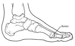
Bunions are readily apparent – the prominence is visible at the base of the big toe or side of the foot. However, to fully evaluate the condition, the foot and ankle surgeon may take x-rays to determine the degree of the deformity and assess the changes that have occurred.
Because bunions are progressive, they don’t go away, and will usually get worse over time. But not all cases are alike – some bunions progress more rapidly than others. Once your surgeon has evaluated your bunion, a treatment plan can be developed that is suited to your needs.
Non-Surgical Treatment
Sometimes observation of the bunion is all that’s needed. To reduce the chance of damage to the joint, periodic evaluation and x-rays by your surgeon are advised.
In many other cases, however, some type of treatment is needed. Early treatments are aimed at easing the pain of bunions, but they won’t reverse the deformity itself. These include:
- Changes in shoewear. Wearing the right kind of shoes is very important. Choose shoes that have a wide toe box and forgo those with pointed toes or high heels which may aggravate the condition.
- Padding. Pads placed over the area of the bunion can help minimize pain. These can be obtained from your surgeon or purchased at a drug store.
- Activity modifications. Avoid activity that causes bunion pain, including standing for long periods of time.
- Medications. Oral nonsteroidal anti-inflammatory drugs (NSAIDs), such as ibuprofen, may be recommended to reduce pain and inflammation.
- Icing. Applying an ice pack several times a day helps reduce inflammation and pain.
- Injection therapy. Although rarely used in bunion treatment, injections of corticosteroids may be useful in treating the inflamed bursa (fluid-filled sac located around a joint) sometimes seen with bunions.
- Orthotic devices. In some cases, custom orthotic devices may be provided by the foot and ankle surgeon.
When Is Surgery Needed?
If non-surgical treatments fail to relieve bunion pain and when the pain of a bunion interferes with daily activities, it’s time to discuss surgical options with a foot and ankle surgeon. Together you can decide if surgery is best for you.
A variety of surgical procedures is available to treat bunions. The procedures are designed to remove the “bump” of bone, correct the changes in the bony structure of the foot, and correct soft tissue changes that may also have occurred. The goal of surgery is the reduction of pain.
In selecting the procedure or combination of procedures for your particular case, the foot and ankle surgeon will take into consideration the extent of your deformity based on the x-ray findings, your age, your activity level, and other factors. The length of the recovery period will vary, depending on the procedure or procedures performed.